Uncovering a Universe of Novel Targets
01
Patient Derived Models
Matched samples sourced from both healthy and diseased tissues to create patient- derived in vitro models


02
SPC Platform
Advanced Computation
Our computer powers high-throughput structural analysis, identifying disease- specific SPC targets with unparalleled precision, automation, and scalability for drug discovery.
Al + Machine Learning
MD Simulations
Molecular Docking

Empirical Structure Data
Proprietary, Ultra Rapid, High-Sensitivity, Mass Spectrometry-Based Structural Proteomics Technology: Structural Proteomics, Mass Spectrometry.
Structural Proteomics
Mass Spectrometry

03
Identification of Conformational Target
Identification of structural features or epitopes that are unique to the disease state. These are often located on surface proteins and are vital for differentiating between diseased and healthy tissues.

04
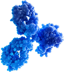
EPIC Platform
Epitope-specific antibody discovery
Our structure-based screening identifies antibodies that bind exclusively to disease- specific protein conformations —sparing normal, healthy protein conformations-for unmatched precision and safety


05
Affinity Optimization
With empirical-data-driven docking and advanced molecular dynamics simulations, we refine discovered antibodies for maximum therapeutic efficacy, stability, and manufacturability. Providing patients with safe, effective and novel therapies.